
respiratory system
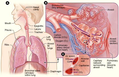
Figure 1. [A] shows the location of the respiratory structures in the body. [B] is an enlarged image of airways, alveoli, and the capillaries. [C] shows the location of gas exchange between the capillaries and alveoli.
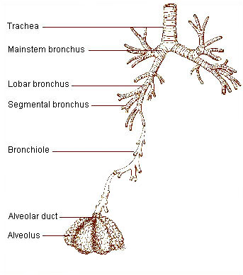
Figure 2. The respiratory tract is divided into two main parts: the upper respiratory tract, consisting of the nose, nasal cavity and the pharynx; and the lower respiratory tract consisting of the larynx, trachea, bronchi and the lungs. The trachea, which begins at the edge of the larynx, divides into two bronchi and continues into the lungs. The trachea allows air to pass from the larynx to the bronchi and then to the lungs. The bronchi divide into smaller bronchioles which branch in the lungs forming passageways for air. The terminal parts of the bronchi are the alveoli. The alveoli are the functional units of the lungs and they form the site of gaseous exchange.
The respiratory system is a group of organs responsible for carrying oxygen from the air to the bloodstream and expelling the waste product carbon dioxide. The main parts of this system are the airways, the lungs and linked blood vessels, and the muscles that enable breathing.
Airways
The airways are passages that carry oxygen-rich air to the lungs and carbon dioxide out of the lungs. The airways include the:
Air first enters the body through the nose or mouth, which wets and warms the air. (Cold, dry air can irritate the lungs.) The air then travels through the larynx and down the trachea. The trachea divides into two bronchi that enter the lungs.
A thin flap of tissue, called the epiglottis, covers the trachea when a person swallows. This prevents food or drink from entering the air passages that lead to the lungs.
Except for the mouth and some parts of the nose, all of the airways have special hairs, called cilia, that are coated with sticky mucus. The cilia trap germs and other foreign particles that enter the airways when a person breathes in air.
These fine hairs then sweep the particles up to the nose or mouth. There, they are swallowed, coughed, or sneezed out of the body. Nose hairs and mouth saliva also trap particles and germs.
Lungs and blood vessels
The lungs and linked blood vessels deliver oxygen to the body and remove carbon dioxide. The lungs lie on either side of the sternum (breastbone) and fill the inside of the chest cavity. The left lung is slightly smaller than the right lung to allow room for the heart.
Within the lungs, the bronchi branch into thousands of smaller, thinner tubes called bronchioles. These tubes end in bunches of tiny round air sacs called alveoli.
Each of these air sacs is covered in a mesh of tiny blood vessels called capillaries. The capillaries connect to a network of arteries and veins that move blood through the body.
The pulmonary artery and its branches deliver blood rich in carbon dioxide (and lacking in oxygen) to the capillaries that surround the air sacs. Inside the air sacs, carbon dioxide moves from the blood into the air. Oxygen moves from the air into the blood in the lungs.
The oxygen-rich blood then travels to the heart through the pulmonary vein and its branches. The heart pumps the oxygen-rich blood out to the body.
The lungs are divided into five main sections called lobes. Some people need to have a diseased lung lobe removed. However, they can still breathe well using the rest of their lung lobes.
Muscles used for breathing
Muscles near the lungs help expand and contract (tighten) the lungs to allow breathing. These muscles include the:
The diaphragm is a dome-shaped muscle located below the lungs. It separates the chest cavity from the abdominal cavity. The diaphragm is the main muscle used for breathing.
The intercostal muscles are located between the ribs. They also play a major role in helping you breathe.
Beneath the diaphragm are abdominal muscles. These help when a person is breathing fast (for example, during physical activity).
Muscles in the neck and collarbone area help a person breathe in when other muscles involved in breathing don't work properly, or when lung disease impairs breathing.
Distinction between respiration and breathing
When the respiratory system is mentioned, people generally think of breathing, but breathing is only one of the activities involved in respiration. The body cells need a continuous supply of oxygen for the metabolic processes that are necessary to maintain life. The respiratory system works with the circulatory system to provide this oxygen and to remove the waste products of metabolism. It also helps to regulate pH of the blood.
Every 3 to 5 seconds, nerve impulses stimulate the breathing process, or ventilation, which, as described above, moves air through a series of passages into and out of the lungs. After this, there is an exchange of gases between the lungs and the blood. This is called external respiration. The blood transports the gases to and from the tissue cells. The exchange of gases between the blood and tissue cells is internal respiration. Finally, the cells utilize the oxygen for their specific activities. This is cellular metabolism, or cellular respiration. Together these activities constitute respiration.
What happens when you breathe?
Breathing in (inhalation)
When a person breathes in, the diaphragm contracts (tightens) and moves downward. This increases the space in the chest cavity, into which the lungs expand. The intercostal muscles between the ribs also help enlarge the chest cavity. They contract to pull the rib cage both upward and outward when the person inhales.
As the lungs expand, air is sucked in through the nose or mouth. The air travels down the windpipe and into the lungs. After passing through the bronchial tubes, the air finally reaches and enters the alveoli (air sacs).
Through very thin walls of the alveoli, oxygen from the air passes to the surrounding capillaries (blood vessels). A red blood cell protein called hemoglobin helps move oxygen from the air sacs to the blood. (Oxygen is especially drawn to hemoglobin.)
At the same time, carbon dioxide moves from the capillaries into the air sacs. The gas has traveled in the bloodstream from the right side of the heart through the pulmonary artery.
Oxygen-rich blood from the lungs is carried through a network of capillaries, which become the pulmonary vein. This vein delivers the oxygen-rich blood to the left side of the heart. The left side of the heart pumps the blood to the rest of the body. There, the oxygen in the blood moves from blood vessels into surrounding tissues.
Breathing out (exhalation)
When a person breathes out, the diaphragm relaxes and moves upward into the chest cavity. The intercostal muscles between the ribs also relax to make the chest cavity size smaller.
As the chest cavity gets smaller, air rich in carbon dioxide is forced out of the lungs and windpipe, and then out of the nose or mouth.
Breathing out requires no effort from the body unless a person has a lung disease or is doing physical activity. When a person is physically active, the abdominal muscles contract and push the diaphragm even more so against the lungs. This pushes the air in the lungs out rapidly.
Lung diseases and conditions
Many steps are involved in breathing. If injury, disease, or other factors affect any of the steps, a person may have trouble breathing.
For example, the fine hairs (cilia) that line the upper airways may not trap all of the germs that are breathed in. These germs can cause an infection in the bronchi (bronchitis) or deep in the lungs (pneumonia). These infections cause a buildup of mucus and/or fluid that narrows the airways and hinders airflow in and out of the lungs.
If a person has asthma, breathing in certain substances to which that individual is sensitive can trigger the airways to narrow. This makes it hard for air to flow in and out of the lungs.
Over a long period, breathing in cigarette smoke or air pollutants can damage the airways and the air sacs. This can lead to a condition called chronic obstructive pulmonary disease (COPD). COPD prevents proper airflow in and out of the lungs and can hinder gas exchange in the air sacs.
An important step to breathing is the movement of the diaphragm and other muscles in the chest, neck, and abdomen. This movement lets a person inhale and exhale. Nerves that run from the brain to these muscles control their movement. Damage to these nerves in the upper spinal cord can cause breathing to stop, unless a machine is used to help in breathing. (This machine is called a ventilator or a respirator.)
A steady flow of blood in the small blood vessels that surround the air sacs is vital for gas exchange. Long periods of inactivity or surgery can cause a blood clot called a pulmonary embolism to block the lung artery. This reduces or stops the flow of blood in the small blood vessels and interferes with gas exchange.

